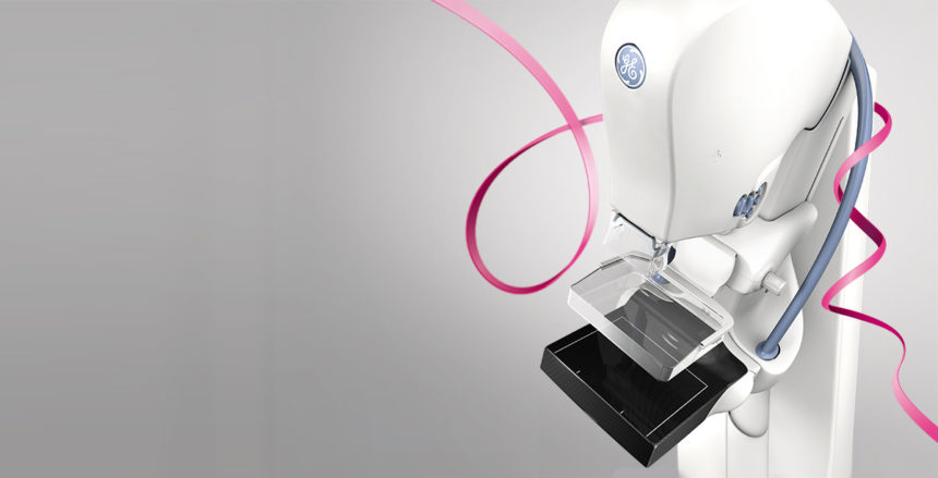
Breast Biopsy
Breast Biopsy & Breast Cancer Diagnosis
Please review our Biopsy FAQ for commonly asked questions about this breast imaging procedure.
We are leaders in the field of the early detection of breast cancer and imaging after a breast cancer diagnosis. We pride ourselves not only on our state-of-the-art imaging facilities, but also on our highly trained and compassionate staff, including our Medical Director, Christopher L. Petti, M.D.
Biopsy Procedures at Bergen Imaging Center
A breast biopsy is a diagnostic procedure in which cells are removed from a suspicious mass on the breast, and examined in order to determine whether they are benign or malignant. Bergen Imaging Center offers ultrasound guided biopsies. During this procedure, your radiologist uses an ultrasound to guide the needle to the biopsy site. This is also referred to as an “image guided” biopsy, and offers greater accuracy in pinpointing the abnormal growth compared to other types of biopsies. We offer three different biopsy procedures including: ultrasound guided fine needle aspirations, ultrasound guided cyst aspirations, and ultrasound guided core needle biopsies.
Ultrasound Guided Fine Needle Aspiration (FNA)
This fast and simple procedure uses ultrasound technology to guide a very fine needle to the area in question (an abnormal growth, lump, etc.). A small liquid tissue sample is then removed and sent to the laboratory for further examination. The process takes around 20-30 minutes.
Ultrasound Guided Cyst Aspiration
This procedure is similar to FNA but is used specifically for the removal or drainage of breast cysts.
Ultrasound Guided Core Needle Biopsy
This procedure uses ultrasound technology to guide a hollow needle to the area in question. The needle is reinserted multiple times in order to obtain solid tissue samples, which are then sent to a laboratory for further examination. This process takes around 30 minutes.
Patients will be called with their laboratory results in 3-5 business days.
FAQ: Ultrasound Guided Breast Biopsy
A breast biopsy is a diagnostic procedure in which cells are removed from a suspicious mass on the breast, and examined in order to determine whether they are benign or malignant. Bergen Imaging Center offers ultrasound guided biopsies. During this procedure, your radiologist uses an ultrasound to guide the needle to the biopsy site. This is also referred to as an “image guided” biopsy, and offers greater accuracy in pinpointing the abnormal growth compared to other types of biopsies.
We offer three different biopsy procedures including: ultrasound guided fine needle aspirations, ultrasound guided cyst aspirations, and ultrasound guided core needle biopsies.
A core needle biopsy is a type of biopsy that uses a needle to obtain small cores of tissue from the lesion in question. After the area is numbed with lidocaine, a core needle is inserted through the skin of the breast. A small core of tissue is removed. Several samples of the lesion will be taken through the same small opening in the skin. This test may cause a bruise, but rarely leaves a scar. The patient can safely drive home after the procedure. The only restrictions are to keep it dry and to avoid heavy lifting or exercising until the next day. The specimen is then sent to the contracted lab for pathologic evaluation and the patient gets results within a few days.
If your doctor finds a lump, bump, or other abnormality on your mammogram, he or she will most likely refer you to a local women’s imaging center for a breast biopsy. While fear and anxiety are natural reactions, rest assured that the vast majority of biopsies – 4 out of 5 – test negative for cancer. In the worst-case scenario, a breast biopsy can save your life by detecting the illness early on.
Ultrasound guided biopsies are faster and less invasive than surgical biopsies. The entire procedure takes under an hour and leaves little or no scarring. Other advantages include:
- No ionizing radiation
- Less expensive than stereotactic or surgical biopsies
- Minimal recovery time required
- Higher accuracy in determining whether a breast abnormality is benign or malignant
- Ability to evaluate lumps in hard to reach places such as under the arm or near the chest
Image-guided, minimally invasive ultrasound guided breast biopsies are performed by a radiologist on an outpatient basis.
You will be positioned face up, or slightly on your side on the examination table, as the area to be biopsied is numbed with anesthetic. Then, using the ultrasound probe to visualize the location of the suspicious mass, your radiologist will insert the biopsy needle into your skin, advance it towards the lump, and extract the necessary tissue samples for examination. Exact details of the procedure vary depending on the type of biopsy performed (ultrasound guided fine needle aspiration, ultrasound guided cyst aspiration, or ultrasound guided core needle biopsy).
While you may experience some bruising, scarring is rare. It is important to keep the biopsy site dry, and avoid exercise or heavy lifting for 24 hours. You will be contacted once your results are in, generally within a few days.
If your biopsy results in a breast cancer diagnosis, it’s important to remember that breast cancer is often highly treatable, especially when detected early. At Bergen Imaging Center, while we focus on diagnostic services, we encourage patients to closely collaborate with their healthcare providers for the next steps.
Your medical team will guide you through understanding your diagnosis, including the type and stage of breast cancer, and discuss various treatment options. Advancements in breast cancer treatments have significantly improved outcomes, and many patients go on to lead healthy, fulfilling lives post-treatment.
Remember, you’re not alone in this journey. Your healthcare provider will provide support and guidance, tailoring a treatment plan to your unique situation. The emphasis will be on effective treatment strategies and supportive care to navigate this challenging phase with positivity and resilience.