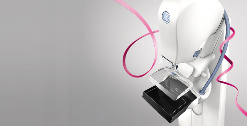
Breast tomosynthesis, more commonly known as 3D mammography, looks and feels a lot like a traditional mammogram. It is performed in conjunction with your 2D mammogram, and does not require additional time or compression of the breasts. *
However, instead of imaging the breast as a whole, 3D mammography provides three-dimensional views by taking multiple X-rays of each breast from different angles. It enables radiologists to view inside of the breast, layer by layer, while increasing visibility of fine details by minimizing the appearance of overlapping tissue.
Research demonstrates that 3D mammography is 30 percent more accurate in finding breast cancer than 2D exams alone – and that it greatly reduces the number of false positive readings. In addition, patients are exposed to very low levels of radiation.
Research presented by the Radiological Society of North America (RSNA), found that tomosynthesis also has potential to significantly increase the cancer detection rate in mammography screening of women with dense breasts. Using digital mammography plus tomosynthesis, researchers detected 80 percent of 132 cancers in women with dense breasts, compared to only 59 percent for mammography alone. Across all breast densities, 82 percent of cancers were detected with 3D mammography, versus 63 percent of cancers being detected using only digital mammography. This is because 3D mammography can “see through” the tissue, making it possible to locate tumors that would otherwise be obscured on a 2D mammogram.
“Tomosynthesis research has also demonstrated a decrease in the number of callbacks, which results in the reduction of additional testing and patient anxiety. Our goal is to make breast tomosynthesis with digital mammography our standard protocol for all women undergoing mammography,” said Christopher L. Petti, MD, Medical Director at Bergen Imaging Center.
* The FDA requires that 3D mammography be performed in conjunction with standard 2D mammography.

